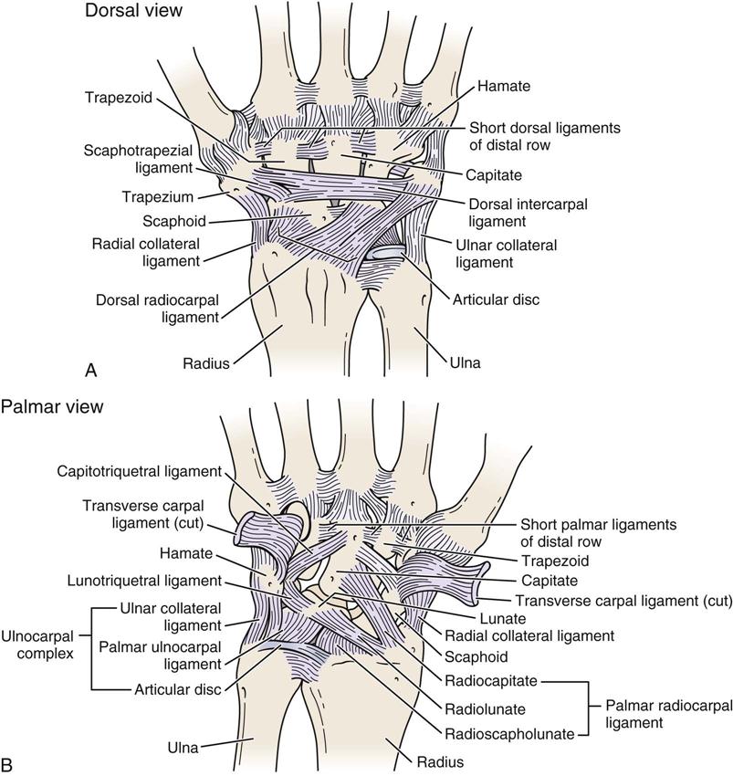
Wrist Ligaments Diagram

Wrist Joint Anatomy Bones Movements Ligaments Tendons Abduction Ulnocarpal and radiocarpal ligaments: ligaments that stabilize your whole wrist while it moves. collateral ligaments: these are the same ligaments as the ones in your hand. they run on both sides on the outside of your wrist and hold your wrist in place. volar carpal ligaments: ligaments that support and stabilize the bottom (palmar side) of. The ligaments of the wrist include. extrinsic ligaments. bridge carpal bones to the radius or metacarpals. include volar and dorsal ligaments. intrinsic ligaments. originate and insert on carpal bones. the most important intrinsic ligaments are the scapholunate interosseous ligament and lunotriquetral interosseous ligament.

Wrist Ligaments Biomechanics Hand Orthobullets The wrist joint (also known as the radiocarpal joint) is an articulation between the radius and the carpal bones of the hand. it is condyloid type synovial joint which marks the area of transition between the forearm and the hand. in this article, we shall look at the anatomy of the wrist joint – its structure, neurovasculature and clinical. Wrist anatomy is the study of the bones, ligaments and other structures in the wrist. the wrist joint is a complex joint which connects the forearm to the hand, allowing a wide range of movement. however, it is susceptible to injury, especially from repetitive strain. advert. The radiocarpal joint is a synovial joint formed between the radius, its articular disc and three proximal carpal bones; the scaphoid, lunate and triquetral bones. technically, the radiocarpal joint is considered to be the only articular component of the wrist joint; many references, however, may also include adjacent joints, such as the carpal. Two important ligaments support the sides of the wrist. these are the collateral ligaments. there are two collateral ligaments that connect the forearm to the wrist, one on each side of the wrist. as its name suggests, the ulnar collateral ligament (ucl) is on the ulnar side of the wrist. it crosses the ulnar edge (the side away from the thumb.

Wrist Ligaments Diagram The radiocarpal joint is a synovial joint formed between the radius, its articular disc and three proximal carpal bones; the scaphoid, lunate and triquetral bones. technically, the radiocarpal joint is considered to be the only articular component of the wrist joint; many references, however, may also include adjacent joints, such as the carpal. Two important ligaments support the sides of the wrist. these are the collateral ligaments. there are two collateral ligaments that connect the forearm to the wrist, one on each side of the wrist. as its name suggests, the ulnar collateral ligament (ucl) is on the ulnar side of the wrist. it crosses the ulnar edge (the side away from the thumb. The wrist joint, also known as the radiocarpal joint, is a crucial connection between the forearm and hand. it allows various movements like bending, straightening, moving side to side, and twisting. this joint is like a modified ball and socket, allowing flexibility while maintaining stability. the wrist anatomy (joint) is made up of bones. Radiocarpal joint. ellipsoid shape involving distal radius and the scaphoid, lunate, and triquetrum. it’s located at the level of the crease of proximal wrist flexion. extrinsic ligaments bridge carpal bones to radius or metacarpals (radioscaphocapitate); while intrinsic attach carpal bones together (scapholunate). carpal bones anatomy.

Comments are closed.