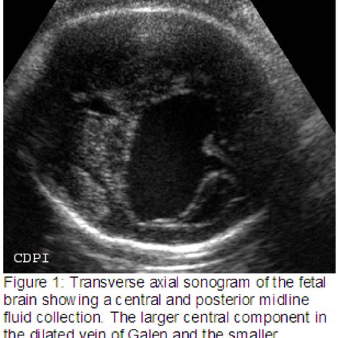
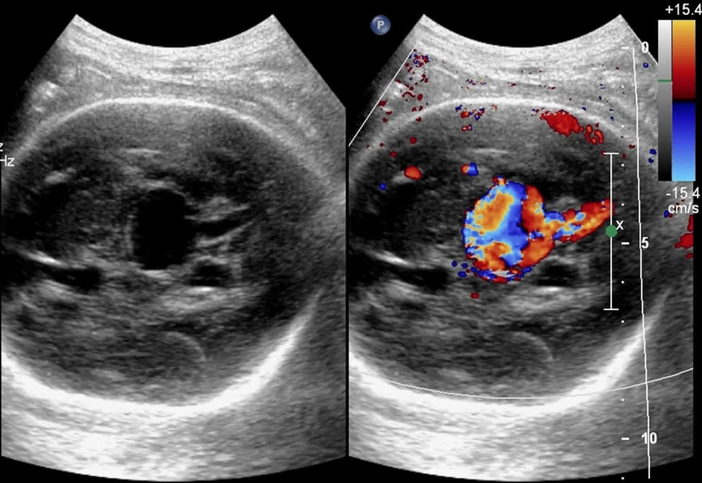
Vein Of Galen Avm Veins Ultrasound Rectangle Glass

Vein Of Galen Avm Veins Ultrasound Rectangle Glass Vrogue Co The mpv fails to regress and becomes aneurysmal. it drains via the straight sinus (present only in 50%) or a persistent falcine sinus, and the vein of galen does not form. haemodynamically cerebral arteriovenous fistula involving vein of galen can be subdivided into two groups: true vgams. vein of galen dilatation secondary to high flow. Termed vein of galen aneurysmal dilatations, these lesions are characterized by drainage of an arteriovenous malformation or dural fistula into the true vein of galen. the dilated vein in these cases drains brain parenchyma in addition to the malformation, as opposed to the persistent embryonic vein in the true vgam that drains only the malformation ( 4 ).

Vein Of Galen Avm Veins Ultrasound Rectangle Glass Vrogue Co Vein of galen aneurysmal malformation (vgam), a rare congenital intracranial arteriovenous (av) malformation (avm) of the cerebral vasculature, represents about 30% of all prenatally diagnosed. Abstract. our approach to treating a patient with a vein of galen aneurysm is, of course, influenced greatly by the age of the patient, the clinical symptoms, and the angiographic architecture of the malformation. therapeutic options are primarily based on whether a true avm is present or if the malformation represents an arteriovenous fistula. The vein of galen aneurysmal malformation (vgam) is a rare arteriovenous malformation of the embryonic choroid plexus. they represent about 30% of all paediatric neurovascular disorders[ 2 , 3 ]. the vgam is constituted by a midline dilated venous structure that receives blood from abnormal macroscopic or microscopic arteriovenous shunting vessels[ 4 ]. Vogm is an embryonic choroid plexus avm, distinct from other deep seated avms with venous drainage into a more mature vein of galen. 3 early in brain development, arterial supply is derived from the choroid plexuses and several associated choroidal arteries while venous drainage primarily occurs through the mpv. 8 in gestational weeks 8 to 11, the mpv segment proximal to its connection with.

Vein Of Galen Avm Veins Ultrasound Rectangle Glass Vrogue Co The vein of galen aneurysmal malformation (vgam) is a rare arteriovenous malformation of the embryonic choroid plexus. they represent about 30% of all paediatric neurovascular disorders[ 2 , 3 ]. the vgam is constituted by a midline dilated venous structure that receives blood from abnormal macroscopic or microscopic arteriovenous shunting vessels[ 4 ]. Vogm is an embryonic choroid plexus avm, distinct from other deep seated avms with venous drainage into a more mature vein of galen. 3 early in brain development, arterial supply is derived from the choroid plexuses and several associated choroidal arteries while venous drainage primarily occurs through the mpv. 8 in gestational weeks 8 to 11, the mpv segment proximal to its connection with. Vein of galen malformation has been associated with capillary malformation arteriovenous malformation (cm avm), which is a newly recognized autosomal dominant disorder, caused by mutations in the rasa1 gene in 6 families. the authors report severe intracranial avms, including vein of galen aneurysmal malformation, which was symptomatic at birth or during infancy, extracranial avm of the face. Vein of galen aneurysmal malformation: an updated review.

Comments are closed.