Restless Nature Of Human Spinal Cord Revealed By Non Invasive Functional Imaging

Restless Nature Of Human Spinal Cord Revealed By Non In Restless nature of human spinal cord revealed by non invasive functional imaging. sciencedaily . retrieved september 2, 2024 from sciencedaily releases 2020 09 200909114846.htm. Restless nature of human spinal cord revealed by non invasive functional imaging. the spinal cord roughly looks like a long tube, with a diameter of only 1.5 cm, and yet this crucial part of the.

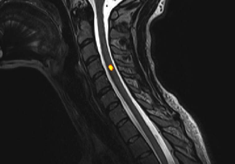
Restless Nature Of Human Spinal Cord Revealed By Non In Restless nature of human spinal cord revealed by non invasive functional imaging september 9 2020 the spinal cord roughly looks like a long tube, with a diameter of only. Epfl scientists have developed a non invasive technique for unraveling the complex dynamics generated by spinal cord circuits to unprecedented detail, a fir. Epfl scientists have developed a non invasive technique for unraveling the complex dynamics generated by spinal cord circuits to unprecedented detail, a first in functional magnetic resonance imaging that may one day help diagnose spinal cord dysfunction or injury. Scientists have longed for a way to observe the spinal cord's function in vivo. until recently, they needed to resort to animal studies, but the advent of fmri (functional magnetic resonance imaging) is now providing a new window into the richness of spinal cord signals, directly in humans.

Restless Nature Of Human Spinal Cord Non Invasive Imagi Epfl scientists have developed a non invasive technique for unraveling the complex dynamics generated by spinal cord circuits to unprecedented detail, a first in functional magnetic resonance imaging that may one day help diagnose spinal cord dysfunction or injury. Scientists have longed for a way to observe the spinal cord's function in vivo. until recently, they needed to resort to animal studies, but the advent of fmri (functional magnetic resonance imaging) is now providing a new window into the richness of spinal cord signals, directly in humans. Stimulation of the spinal cord can be achieved using non invasive methodology whereby electrical current is delivered to the spinal cord through surface electrodes, so as to modulate neuronal. Agyeman et al. leverage functional ultrasound imaging (fusi) to characterize human spinal cord activity during epidural electrical stimulation. they decode the stimulation’s effectiveness to evoke hemodynamic changes at the single trial level. this establishes fusi as a promising platform for investigating spinal cord function and developing real time closed loop clinical neuromodulation.

Comments are closed.