Mri Pulse Sequence Diagrams Heart Failure Guws Medical Images

Mri Pulse Sequence Diagrams Heart Failure Guws Medica Vrog The second gradient pulse, of opposite polarity, can reverse this effect. an echo type signal is observed and peaks at the point at which the phase warp produced by the first pulse is cancelled. this type of echo is called a gradient echo. a pulse sequence diagram for a gradient echo imaging sequence is shown in fig. 8. Objective. mri is a well established modality for evaluating congenital and acquired cardiac diseases. this article reviews the latest pulse sequences used for cardiac mri. in addition, the standard cardiac imaging planes and corresponding anatomy are described and illustrated. conclusion. familiarity with the basic pulse sequences, imaging planes, and anatomy pertaining to cardiac mri is.

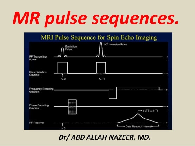
Mri Pulse Sequence Diagrams Heart Failure Guws Medical Images Annotations #. in most every mri experiment, the basic pulse sequence block is repeated but with a different gradient pattern to perform the necessary spatial encoding. this is typically annotated in 2 ways. first, the changes in gradient patterns can be added through multiple lines on the gradient axes. second, bracketing around the sequence. Published online: december 21, 2023. understanding the fundamentals of body mri pulse sequences, including the protocol framework, tools and techniques, sequence families, quantitative imaging, motion reduction, and protocol design, will aid radiologists in maximizing diagnostic yield. download as powerpoint ». Figure 4.2. pulse sequence diagrams for the 4 basic pulse sequence types used in cardiac mr imaging: spin echo, multiecho spin echo (or multiple spin echo), spoiled gradient echo, and balanced gradient echo. each sequence shows the rf pulse (s) and spatial encoding gradients used to create a single image echo. May 3 0, 2010. cardiac imaging. objective. mri is a well established modality for evaluating congenital and acquired car. diac disease s. this ar ticle reviews the latest pulse sequences used for.

Mri Pulse Sequence Diagrams Heart Failure Guws Medica Vrog Figure 4.2. pulse sequence diagrams for the 4 basic pulse sequence types used in cardiac mr imaging: spin echo, multiecho spin echo (or multiple spin echo), spoiled gradient echo, and balanced gradient echo. each sequence shows the rf pulse (s) and spatial encoding gradients used to create a single image echo. May 3 0, 2010. cardiac imaging. objective. mri is a well established modality for evaluating congenital and acquired car. diac disease s. this ar ticle reviews the latest pulse sequences used for. Multiple sequences are usually needed to adequately evaluate a tissue, and the combination of sequences is referred to as a mri protocol. the radiologist tailors the pulse sequences to try to best answer the clinical question posed by referring physician. protocols are discussed more fully in a separate article: mri protocols. Cardiac mri pulse sequences. pulse sequences are software programs that encode the magnitude and timing of the radiofrequency pulses emitted by the mr scanner, switching of the magnetic field gra dient, and data acquisition. the components of a pulse sequence are termed “imaging en gines” and “modifiers” [8].

Comments are closed.