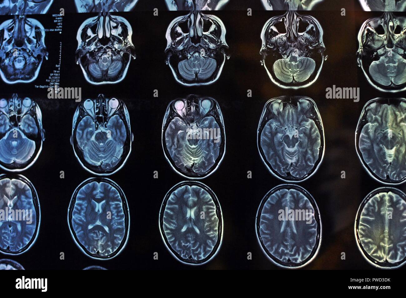
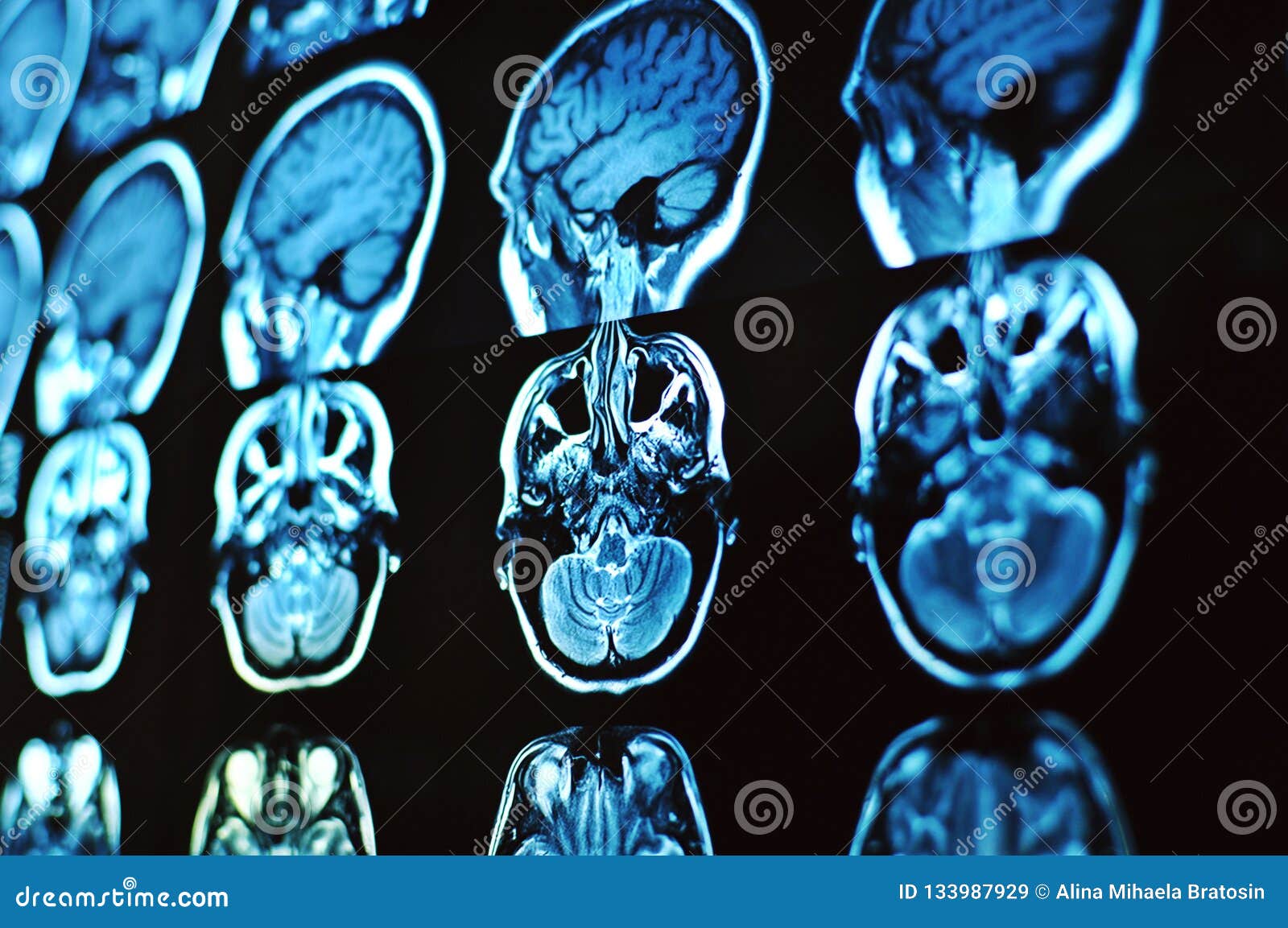
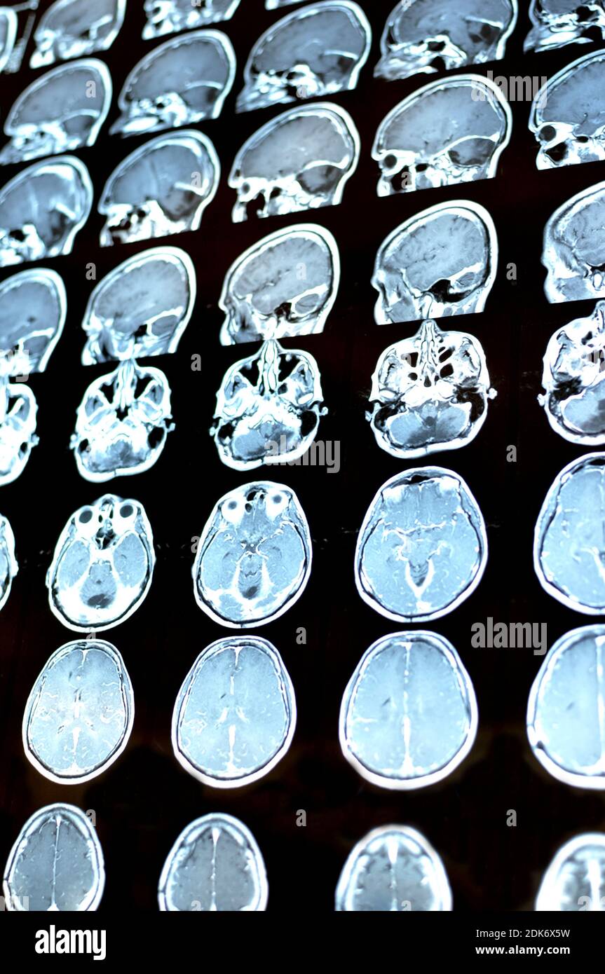
Magnetic Resonance Image Scan Of The Brain Mri Film Of A Human Skull

Magnetic Resonance Image Scan Of The Brain Mri Film Of A Human Skull A brain mri (magnetic resonance imaging) scan, also called a head mri, is a painless procedure that produces very clear images of the structures inside of your head — mainly, your brain. mri uses a large magnet, radio waves and a computer to produce these detailed images. it doesn’t use radiation. currently, mri is the most sensitive. An mri machine creates the images using a magnetic field and radio waves. this test is also known as a brain mri or a cranial mri. you will go to a hospital or radiology center to take a head mri.

Magnetic Resonance Image Scan Of The Brain Mri Film Of A Human Skull Brain mri. a brain mri, also called a head mri, uses a powerful magnetic field, radio waves and a computer to produce pictures of the brain. the pictures produced are clearer and more detailed than other imaging methods. magnetic resonance imaging (mri) does not use ionizing radiation and may require an injection of a contrast material called. Ops 301 code. 3 800, 3 820. magnetic resonance imaging of the brain uses magnetic resonance imaging (mri) to produce high quality two dimensional or three dimensional images of the brain and brainstem as well as the cerebellum without the use of ionizing radiation (x rays) or radioactive tracers. Magnetic resonance imaging (mri) is a noninvasive technique used for diagnostic imaging. mri is particularly useful for the imaging of soft tissues. therefore, mri allows for high quality imaging of the brain with good anatomic detail and offers more sensitivity and specificity than other imaging modalities for many types of neurologic conditions. Learning that you need to undergo a magnetic resonance imaging (mri) test can be intimidating. an mri of the brain can be used to evaluate many symptoms which may be caused by abnormalities in the central nervous system. these include headache, seizures, sleep disorders, mental confusion, weakness, numbness, or dizziness.

Magnetic Resonance Image Scan Of The Brain With A Colloid Cyst Mriо Magnetic resonance imaging (mri) is a noninvasive technique used for diagnostic imaging. mri is particularly useful for the imaging of soft tissues. therefore, mri allows for high quality imaging of the brain with good anatomic detail and offers more sensitivity and specificity than other imaging modalities for many types of neurologic conditions. Learning that you need to undergo a magnetic resonance imaging (mri) test can be intimidating. an mri of the brain can be used to evaluate many symptoms which may be caused by abnormalities in the central nervous system. these include headache, seizures, sleep disorders, mental confusion, weakness, numbness, or dizziness. Magnetic resonance imaging (mri) is a non invasive imaging technology that produces three dimensional detailed anatomical images. it is often used for disease detection, diagnosis, and treatment monitoring. it is based on sophisticated technology that excites and detects the change in the direction of the rotational axis of protons found in the. Magnetic resonance imaging (mri) is a diagnostic procedure that uses a combination of a large magnet, radiofrequencies, and a computer to produce detailed images of organs and structures within the body. unlike x rays or computed tomography (ct scans), mri does not use ionizing radiation. some mri machines look like narrow tunnels, while others.

Comments are closed.