Internal Cerebral Vein Neuroangio Org

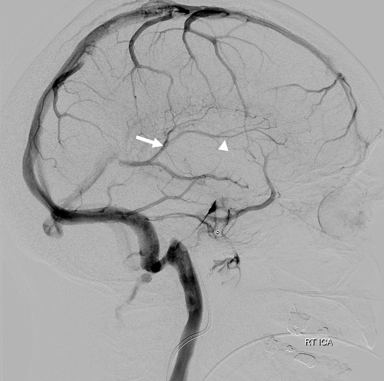
Internal Cerebral Vein Neuroangio Org The best way to understand internal cerebral vein anatomy (and, actually, any venous anatomy) is to: 1 — study how the same veins appear on cross sectional imaging and angiography. in particular it is important to understand the ventricles. 2 — understand the concept of concentric rings (see below) and balance between deep and superficial. The “superficial” — meaning surface — veins are generally seen first. this includes the basal vein, which is in effect a superficial (surface) vein that happens to be on the bottom of the brain. from here, contrast progresses into the venous sinuses, which are usually opacified well about 1 1.5 seconds after the superficial cortical veins.

Internal Cerebral Vein Neuroangio Org Neuroangio.org has gone bananaz. in response to the global shift of education to online platforms, neuroangio.org is providing free online neurovascular anatomy education via our bananaz monthly seminars. the author, maksim shapiro, md is a neurointerventional radiologist in at the nyu langone medical center in new york city, and can be reached. Figure 2 (a–l) internal cerebral, basal, and deep venous systems (a–l), highlighting venous drainage balance. (a) the idea of “concentric rings” or grid like arrangement can be very helpful in conceptualizing the deep system and its variations internal cerebral vein – pink; basal – black; thalamostriate – brown; septal – white; direct lateral – yellow; longitudinal caudate. The internal cerebral veins are two veins included in the group of deep cerebral veins that drain the deep parts of the hemispheres; each internal cerebral vein is formed near the interventricular foramina by the union of the superior thalamostriate vein and the superior choroid vein. they run backward parallel with one another, between the. The internal cerebral veins unite with the basal veins (of rosenthal) to form the great cerebral vein (of galen) just beneath the splenium of the corpus callosum in the quadrigeminal cistern. the confluence of the great cerebral vein and inferior sagittal sinus forms the straight sinus. the drainage territory is highly variable and usually.

Internal Cerebral Vein Neuroangio Org The internal cerebral veins are two veins included in the group of deep cerebral veins that drain the deep parts of the hemispheres; each internal cerebral vein is formed near the interventricular foramina by the union of the superior thalamostriate vein and the superior choroid vein. they run backward parallel with one another, between the. The internal cerebral veins unite with the basal veins (of rosenthal) to form the great cerebral vein (of galen) just beneath the splenium of the corpus callosum in the quadrigeminal cistern. the confluence of the great cerebral vein and inferior sagittal sinus forms the straight sinus. the drainage territory is highly variable and usually. Figure 1. venous drainage routes of the brainstem and cerebellum.there are 3 basic drainage routes for the brainstem and cerebellum: (1) anterior drainage (orange arrow, to the superior petrosal sinus [sps]); (2) superior drainage (light blue arrow to the vein of galen [vog]); and (3) posterior drainage (green arrow, to the transverse sinus [ts] and torcular herophili). Indications. the indications for neuroangiography (na) include intracranial diseases (cerebral aneurysms, arteriovenous malformations, dural arteriovenous fistula, cerebral vasculitis, cerebral vasospasm, ischemic stroke, nontraumatic subarachnoid hemorrhage, intracerebral hemorrhage, moyamoya, vein of galen malformation, intracranial tumors, and pseudotumor cerebri) and extracranial diseases.

Internal Cerebral Vein Neuroangio Org Figure 1. venous drainage routes of the brainstem and cerebellum.there are 3 basic drainage routes for the brainstem and cerebellum: (1) anterior drainage (orange arrow, to the superior petrosal sinus [sps]); (2) superior drainage (light blue arrow to the vein of galen [vog]); and (3) posterior drainage (green arrow, to the transverse sinus [ts] and torcular herophili). Indications. the indications for neuroangiography (na) include intracranial diseases (cerebral aneurysms, arteriovenous malformations, dural arteriovenous fistula, cerebral vasculitis, cerebral vasospasm, ischemic stroke, nontraumatic subarachnoid hemorrhage, intracerebral hemorrhage, moyamoya, vein of galen malformation, intracranial tumors, and pseudotumor cerebri) and extracranial diseases.

Comments are closed.