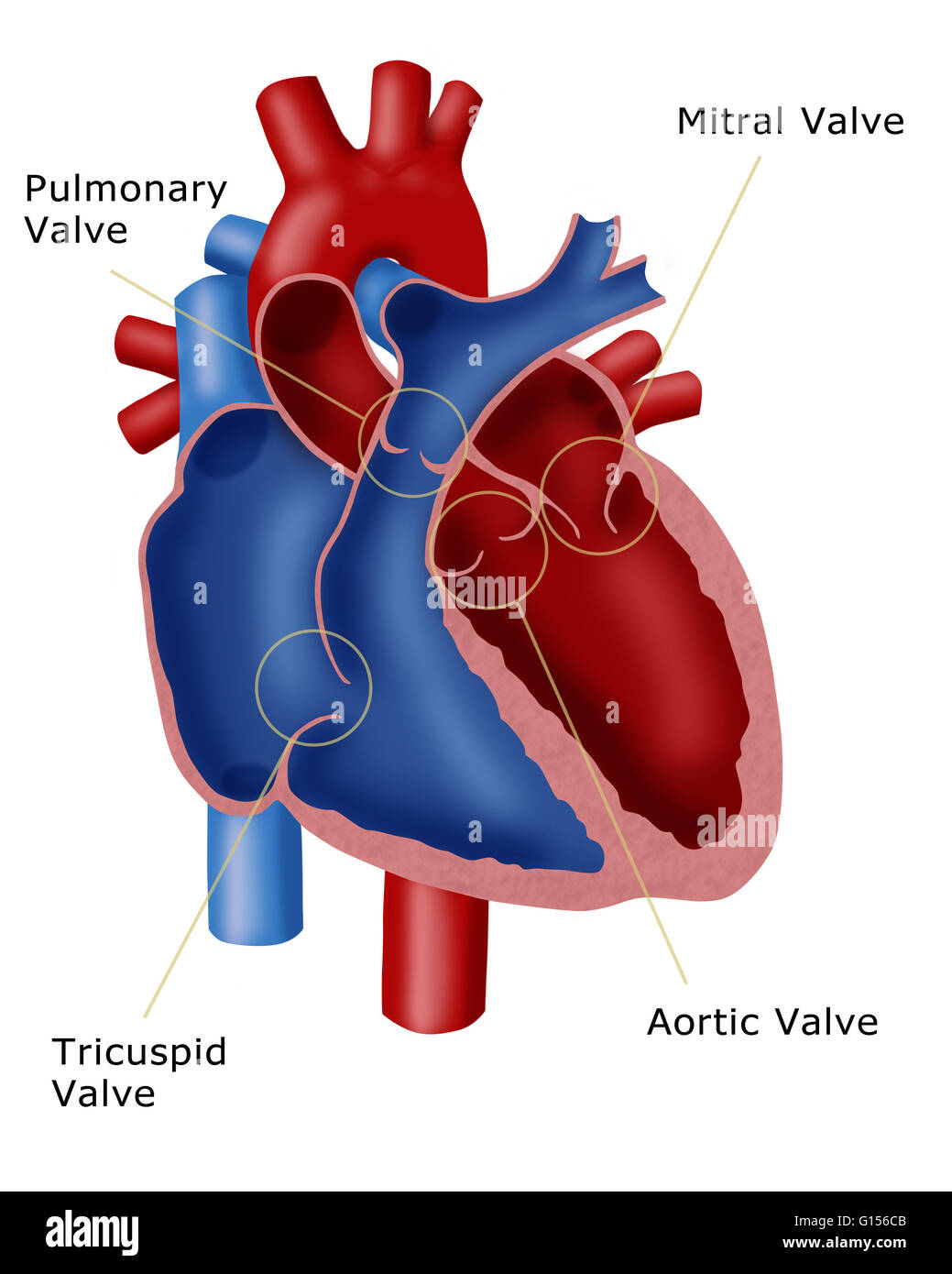
Illustration Of A Heart Showing The Four Valves Pulmonary Valve

Illustration Of A Heart Showing The Four Valves Pulmonary Valve As your heart pumps blood, four valves open and close to make sure blood flows in the correct direction. as they open and close, they make two sounds that create the sound of a heartbeat. the four valves are the aortic valve, mitral valve, pulmonary valve and tricuspid valve. a heart murmur is often the first sign of a heart valve problem. The four heart valves are: the mitral valve. the aortic valve. the tricuspid valve. the pulmonic valve. doctors call the mitral and tricuspid valves the atrioventricular valves, and the aortic and.

Functionality Of Heart Valves Abbott The four valves in order of circulation are: tricuspid valve. has three leaflets or cusps. separates the top right chamber (right atrium) from the bottom right chamber (right ventricle). opens to allow blood to flow from the right atrium to the right ventricle. prevents the back flow of blood from the right ventricle to the right atrium. There are 4 valves of the heart, 2 on the left side and 2 on the right side. the left side of the heart can be thought of as the hearts powerful engine. each side of the heart has a collecting chamber on top (atrium), and a pumping chamber on the bottom (ventricle). there is a valve between the upper and lower chamber, and there is a valve. Right atrioventricular valve. valva atrioventricularis dextra. 1 4. synonyms: tricuspid valve, valva tricuspidalis. understanding heart valves anatomy is important in grasping the overall function of the heart. the heart is one of the most important organs in the body. it is responsible for propelling blood to every organ system, including itself. The valves of the heart are structures which ensure blood flows in only one direction. they are composed of connective tissue and endocardium (the inner layer of the heart). there are four valves of the heart, which are divided into two categories: atrioventricular valves: the tricuspid valve and mitral (bicuspid) valve. they are located.

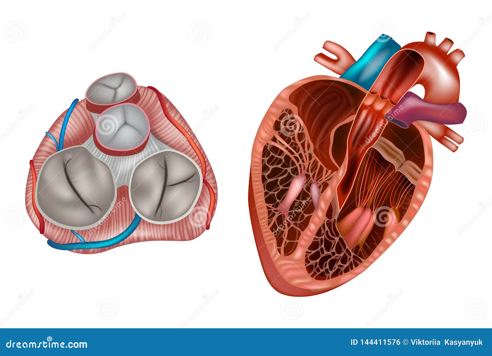
Heart Valves Anatomy Stock Illustration Illustration Of Organ 144411576 Right atrioventricular valve. valva atrioventricularis dextra. 1 4. synonyms: tricuspid valve, valva tricuspidalis. understanding heart valves anatomy is important in grasping the overall function of the heart. the heart is one of the most important organs in the body. it is responsible for propelling blood to every organ system, including itself. The valves of the heart are structures which ensure blood flows in only one direction. they are composed of connective tissue and endocardium (the inner layer of the heart). there are four valves of the heart, which are divided into two categories: atrioventricular valves: the tricuspid valve and mitral (bicuspid) valve. they are located. The heart consists of four chambers, two atria (upper chambers) and two ventricles (lower chambers). there is a valve through which blood passes before leaving each chamber of the heart. the valves prevent the backward flow of blood. these valves are actual flaps that are located on each end of the two ventricles (lower chambers of the heart). The heart is a four chambered organ hemodynamically functioning as a reservoir and a pump; it receives deoxygenated blood from the systemic circulation through the superior and inferior vena cava in the right atrium and oxygenated blood from the lung via the four pulmonary veins. the heart pumps deoxygenated blood from the right ventricle to the lungs and at the same time pumps oxygenated.

Comments are closed.