Heart Diagram With Valves
.svg/1024px-Diagram_of_the_human_heart_(valves_improved).svg.png)
File Diagram Of The Human Heart Valves Improved Svg Wikipedia As your heart pumps blood, four valves open and close to make sure blood flows in the correct direction. as they open and close, they make two sounds that create the sound of a heartbeat. the four valves are the aortic valve, mitral valve, pulmonary valve and tricuspid valve. a heart murmur is often the first sign of a heart valve problem. Learn about the four heart valves that ensure blood flows in the right direction and prevent backward flow. find out about the common problems, symptoms, and treatments of heart valve disorders.
/human-heart-circulatory-system-598167278-5c48d4d2c9e77c0001a577d4.jpg)
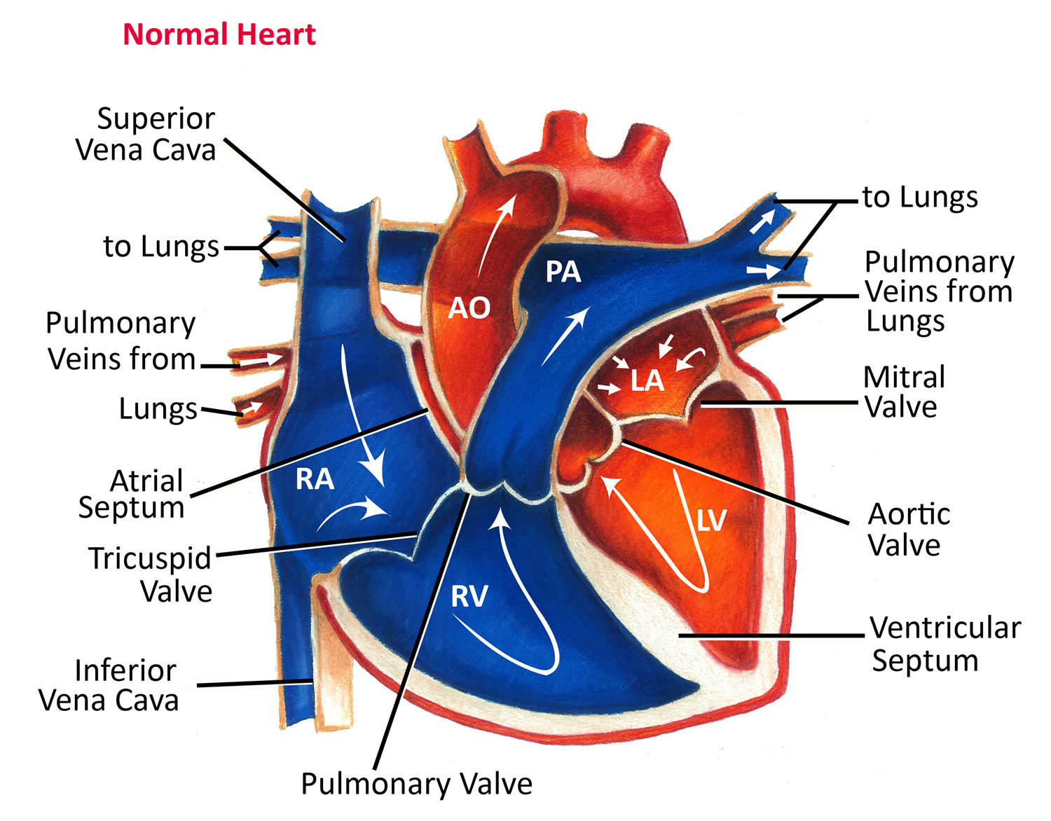
Av And Semilunar Heart Valves Right atrioventricular valve. valva atrioventricularis dextra. 1 4. synonyms: tricuspid valve, valva tricuspidalis. understanding heart valves anatomy is important in grasping the overall function of the heart. the heart is one of the most important organs in the body. it is responsible for propelling blood to every organ system, including itself. They are composed of connective tissue and endocardium (the inner layer of the heart). there are four valves of the heart, which are divided into two categories: atrioventricular valves: the tricuspid valve and mitral (bicuspid) valve. they are located between the atria and corresponding ventricle. semilunar valves: the pulmonary valve and. The aortic semilunar valve is between the left ventricle and the opening of the aorta. it has three semilunar cusps leaflets: left left coronary, right right coronary, and posterior non coronary. in clinical practice, the heart valves can be auscultated, usually by using a stethoscope. Learn about the heart's chambers, valves, and blood flow with 3d models and interactive diagrams. see how the heart wall, atria, ventricles, and cardiac cycle work together to pump blood throughout the body.

Heart Valve Anatomy Britannica The aortic semilunar valve is between the left ventricle and the opening of the aorta. it has three semilunar cusps leaflets: left left coronary, right right coronary, and posterior non coronary. in clinical practice, the heart valves can be auscultated, usually by using a stethoscope. Learn about the heart's chambers, valves, and blood flow with 3d models and interactive diagrams. see how the heart wall, atria, ventricles, and cardiac cycle work together to pump blood throughout the body. Arrhythmias: abnormal heart rhythms, like fast, slow, or irregular beats, can disrupt heart function. the most common type is atrial fibrillation, which causes a fast and irregular heartbeat. heart valve diseases: problems with heart valves can cause inefficient blood flow, leading to symptoms like fatigue, shortness of breath, and dizziness. The four valves in order of circulation are: tricuspid valve. has three leaflets or cusps. separates the top right chamber (right atrium) from the bottom right chamber (right ventricle). opens to allow blood to flow from the right atrium to the right ventricle. prevents the back flow of blood from the right ventricle to the right atrium.

Heart Valves Function Purpose And How Many Heart Valves In Your Heart Arrhythmias: abnormal heart rhythms, like fast, slow, or irregular beats, can disrupt heart function. the most common type is atrial fibrillation, which causes a fast and irregular heartbeat. heart valve diseases: problems with heart valves can cause inefficient blood flow, leading to symptoms like fatigue, shortness of breath, and dizziness. The four valves in order of circulation are: tricuspid valve. has three leaflets or cusps. separates the top right chamber (right atrium) from the bottom right chamber (right ventricle). opens to allow blood to flow from the right atrium to the right ventricle. prevents the back flow of blood from the right ventricle to the right atrium.

Comments are closed.