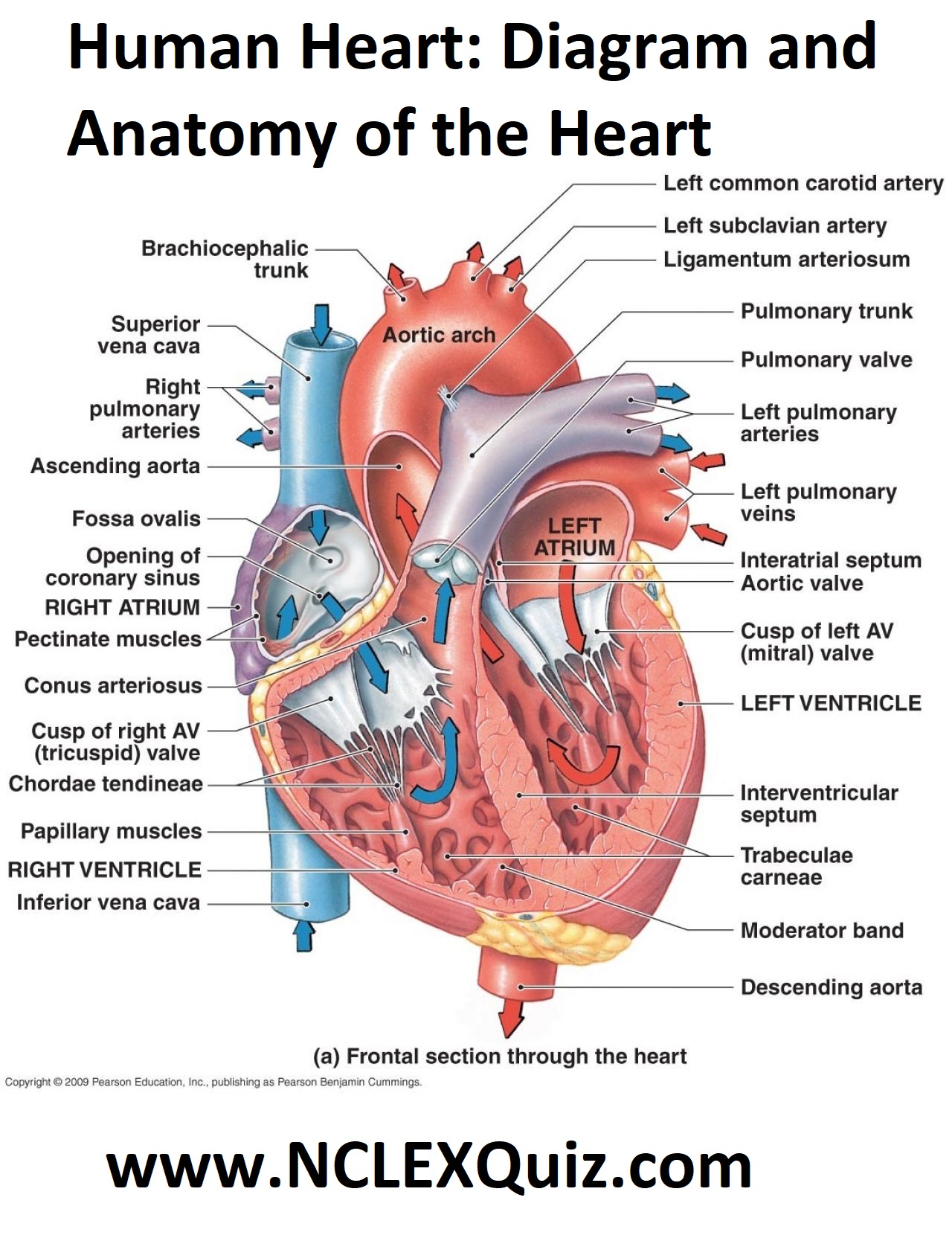
Diagram Of The Inside Of The Heart

Human Heart Diagram And Anatomy Of The Heart Studypk The heart consists of several layers of a tough muscular wall, the myocardium. a thin layer of tissue, the pericardium, covers the outside, and another layer, the endocardium, lines the inside. the heart cavity is divided down the middle into a right and a left heart, which in turn are subdivided into two chambers. Heart anatomy in basic terms. the heart is a crucial organ that functions as the body's pump, ensuring blood circulation throughout the body. it consists of four main chambers: left and right atria (upper chambers) left and right ventricles (lower chambers) these chambers work in a coordinated manner to receive oxygen poor blood, pump it to the.

Interior View Of The Human Heart Medicinebtg The heart has three layers. they are the: epicardium: this thin membrane is the outer most layer of the heart. myocardium: this thick layer is the muscle that contracts to pump and propel blood. The base of the heart is located along the body's midline with the apex pointing toward the left side. because the heart points to the left, about 2 3 of the heart's mass is found on the left side of the body and the other 1 3 is on the right. anatomy of the heart pericardium. the heart sits within a fluid filled cavity called the pericardial. The aortic semilunar valve is between the left ventricle and the opening of the aorta. it has three semilunar cusps leaflets: left left coronary, right right coronary, and posterior non coronary. in clinical practice, the heart valves can be auscultated, usually by using a stethoscope. Muscle and tissue make up this powerhouse organ. your heart contains four muscular sections (chambers) that briefly hold blood before moving it. electrical impulses make your heart beat, moving blood through these chambers. your brain and nervous system direct your heart’s function. advertisement.

Heart Anatomy Part Of The Human Heart Stock Vector Image Art Alamy The aortic semilunar valve is between the left ventricle and the opening of the aorta. it has three semilunar cusps leaflets: left left coronary, right right coronary, and posterior non coronary. in clinical practice, the heart valves can be auscultated, usually by using a stethoscope. Muscle and tissue make up this powerhouse organ. your heart contains four muscular sections (chambers) that briefly hold blood before moving it. electrical impulses make your heart beat, moving blood through these chambers. your brain and nervous system direct your heart’s function. advertisement. Anatomy of the interior of the heart. this image shows the four chambers of the heart and the direction that blood flows through the heart. oxygen poor blood, shown in blue purple, flows into the heart and is pumped out to the lungs. then oxygen rich blood, shown in red, is pumped out to the rest of the body, with the help of the heart valves. 1. the heart wall is composed of three layers. the muscular wall of the heart has three layers. the outermost layer is the epicardium (or visceral pericardium). the epicardium covers the heart, wraps around the roots of the great blood vessels, and adheres the heart wall to a protective sac. the middle layer is the myocardium.
:max_bytes(150000):strip_icc()/heart_interior-570555cf3df78c7d9e908901.jpg)
The 3 Layers Of The Heart Wall Anatomy of the interior of the heart. this image shows the four chambers of the heart and the direction that blood flows through the heart. oxygen poor blood, shown in blue purple, flows into the heart and is pumped out to the lungs. then oxygen rich blood, shown in red, is pumped out to the rest of the body, with the help of the heart valves. 1. the heart wall is composed of three layers. the muscular wall of the heart has three layers. the outermost layer is the epicardium (or visceral pericardium). the epicardium covers the heart, wraps around the roots of the great blood vessels, and adheres the heart wall to a protective sac. the middle layer is the myocardium.

Interior Of The Heart Diagram Medicinebtg

Comments are closed.