Action Of Mylohyoid Muscle

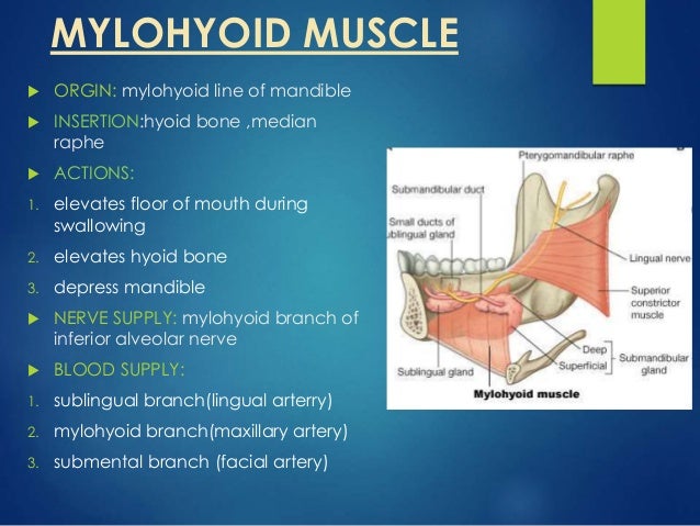
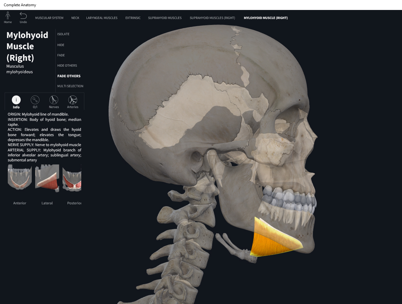
Mylohyoid Muscle Anatomy Wikitomy Mylohyoid muscle in situ: relations with head and neck structures. forming the floor of the mouth, the superior surface of mylohyoid muscle is related to the structures of the oral cavity; it lies directly beneath the geniohyoid, hyoglossus and styloglossus muscles, hypoglossal (cn xii) and lingual nerves, submandibular ganglion, sublingual and submandibular glands, and the lingual artery and. Description. the mylohyoideus (mylohyoid muscle), flat and triangular, is situated immediately above the anterior belly of the digastricus, and forms, with its fellow of the opposite side, a muscular floor for the cavity of the mouth. it arises from the whole length of the mylohyoid line of the mandible, extending from the symphysis in front to.

Anatomy Of Oral Cavity Tongue And Palate The mylohyoid is a suprahyoid muscle of the neck. it is a triangular shaped muscle which forms the floor of the oral cavity and supports the floor of the mouth. attachments: originates from the mylohyoid line of the mandible, and attaches onto the hyoid bone. actions: elevates the hyoid bone and the floor of the mouth. The mylohyoid muscle maintains a critical participant in day to day activities, including mastication and swallowing of food and production of speech in conversations. a key mechanism of action of the mylohyoid muscle is it works directly and indirectly with the infrahyoid muscle to guide the position of the hyoid bone. The mylohyoid muscle is flat and triangular, and is situated immediately superior to the anterior belly of the digastric muscle. it is a pharyngeal muscle (derived from the first pharyngeal arch) and classified as one of the suprahyoid muscles. together, the paired mylohyoid muscles form a muscular floor for the oral cavity of the mouth. The mylohyoid muscle is a thin, flat muscle with a triangular shape that is located above the front part of the digastric muscle. it is a muscle of the pharynx (derived from the first pharyngeal arch) and is classified as one of the suprahyoid muscles. the mylohyoid muscle is made up of two pairs of muscles, which together form a muscular floor.

Mylohyoid Muscle Provides The Blood Supply Of Lower Medial Aspect Of The mylohyoid muscle is flat and triangular, and is situated immediately superior to the anterior belly of the digastric muscle. it is a pharyngeal muscle (derived from the first pharyngeal arch) and classified as one of the suprahyoid muscles. together, the paired mylohyoid muscles form a muscular floor for the oral cavity of the mouth. The mylohyoid muscle is a thin, flat muscle with a triangular shape that is located above the front part of the digastric muscle. it is a muscle of the pharynx (derived from the first pharyngeal arch) and is classified as one of the suprahyoid muscles. the mylohyoid muscle is made up of two pairs of muscles, which together form a muscular floor. The mylohyoid muscles form the muscular floor of the oral cavity. thus, it acts as a diaphragm between the oral cavity and the neck. the inferior (external) aspect of the mylohyoid muscle is related to the anterior belly of digastric, as well as the facial artery and vein. the superior (internal) aspect of the mylohyoid muscle is related to. Abstract. the mylohyoid is one of the suprahyoid muscles, along with the geniohyoid, digastric, and stylohyoid muscles. it lies between the anterior belly of the digastric muscle inferiorly and the geniohyoid superiorly. in part i, the anatomy and embryology of the mylohyoid muscle will be reviewed in preparation for the clinical discussion in.

Muscles Mylohyoid вђ Anatomy Physiology The mylohyoid muscles form the muscular floor of the oral cavity. thus, it acts as a diaphragm between the oral cavity and the neck. the inferior (external) aspect of the mylohyoid muscle is related to the anterior belly of digastric, as well as the facial artery and vein. the superior (internal) aspect of the mylohyoid muscle is related to. Abstract. the mylohyoid is one of the suprahyoid muscles, along with the geniohyoid, digastric, and stylohyoid muscles. it lies between the anterior belly of the digastric muscle inferiorly and the geniohyoid superiorly. in part i, the anatomy and embryology of the mylohyoid muscle will be reviewed in preparation for the clinical discussion in.

Mylohyoid Muscle Attachments Function Human Anatomy Kenhub

Comments are closed.